Glaucoma is a progressive eye condition that affects over 3 million people in the United States. As it progresses, it causes damage to the optic nerve, which can lead to blurred vision or vision loss.
The best way to detect it early is to go for regular eye exams.
Glaucoma can affect anyone, but those with a family history of the disease have a higher risk of developing it. Their likelihood of getting glaucoma is 4–9 times higher than those without a family history.
What Is Glaucoma?
Glaucoma is a group of eye conditions that can cause damage to the optic nerve, leading to vision loss and even blindness. It occurs when the pressure inside the eye (intraocular pressure or IOP) becomes too high. This high pressure causes damage to the optic nerve—the part of your visual system that carries visual information from the eye to the brain.
While glaucoma is more commonly found in adults over 60, it can still affect people of all ages, from infants to seniors. That’s why it’s important for everyone to have their eyes checked regularly.
Risk Factors
A few factors can put you at a higher risk of developing glaucoma. These include:
- Having a family history of the condition
- Being over the age of 60
- Being nearsighted
- Having high intraocular pressure
- Having low blood pressure
If you’re aware of these risk factors, it’s important to bring them up with your eye doctor during your regular exams.
Types of Glaucoma
There are two main types of glaucoma: open-angle and closed-angle.
Open-Angle Glaucoma
Open-angle glaucoma is the most common form, and it occurs when the normal drainage outflow mechanism in your eye gets blocked, leading to increased pressure inside your eye. This type of glaucoma is usually asymptomatic until the optic nerve is severely damaged in the later stages.
At that point, you might experience the following:
- Blind spots in one or both eyes
- Tunnel vision
- Blindness
Some people might have normal-tension glaucoma, a type of open-angle glaucoma where the optic nerve is damaged even when the intraocular pressure isn’t elevated.
Closed-Angle Glaucoma
Closed-angle glaucoma, on the other hand, occurs when your iris is very close to the drainage angle in your eye and can block it, leading to a rapid increase in eye pressure. It can be classified as either acute or chronic.
- Acute closed-angle glaucoma develops quickly and can seriously affect your vision and health. Symptoms might include severe headaches, pain in one or both eyes, nausea or vomiting, blurry vision, halo effects around lights, and red eyes.
- Chronic closed-angle glaucoma develops gradually, making it more difficult to detect. It’s more common in people over 40, farsighted, and women with a family history of the disease.

Detecting Glaucoma
So how is glaucoma detected? Here are a few common methods:
- Eye exams
- Visual field testing
- Optical coherence tomography (OCT)
Eye Exams
The first step in detecting glaucoma is a comprehensive eye exam. During this exam, your eye doctor will check your vision, measure your intraocular pressure (IOP), and examine the health of your eyes. They may also use special tests to assess the health of your optic nerve and measure the thickness of your cornea.
Visual Field Testing
Another way to detect glaucoma is through visual field testing. This test measures your peripheral vision, which can be affected by glaucoma.
During the test, you’ll sit in front of a machine and look straight ahead while lights flash on different parts of the screen. You’ll press a button every time you see a light. This test helps your eye doctor determine if you have any blind spots or areas of decreased vision.
Optical Coherence Tomography (OCT)
An OCT scan is a non-invasive test that uses light waves to create detailed images of the layers of your eye. It’s often used to diagnose and monitor glaucoma because it can show your eye doctor the thickness of your optic nerve and retina (the light-sensitive layer at the back of the eye).
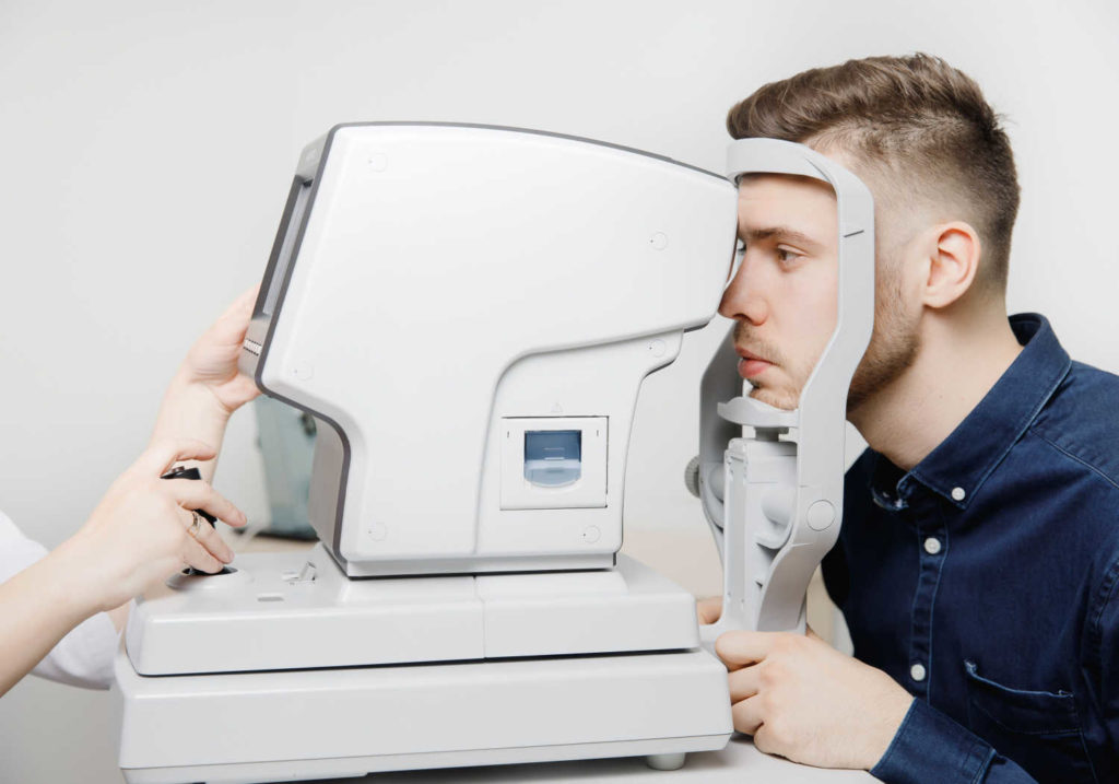
During an OCT test, you will look into a device that shines a light into the eye. The light is reflected off the layers of the retina and captured by a camera, which creates a detailed image of the retina.
These images can help the eye doctor detect any abnormalities or changes in the retina, such as thinning or swelling of the layers, which can signify a potential eye problem. It can also help them see if there are any changes in the nerve fibers that carry visual information from the eye to the brain.
Treating Glaucoma at Perspective Eye Center
The main goal of glaucoma treatment is to lower the pressure in the eye to prevent further damage to the optic nerve. This can be done with medications, such as eye drops or pills, or surgery.If it’s time for your next eye exam or you have more questions about glaucoma and its risk factors, book an appointment with our team at Perspective Eye Center.


