
Your optometrist conducts several tests during a comprehensive eye exam. One of the ways they check for eye diseases is with an optical coherence tomography test.
What is this diagnostic test, and how does it work? Continue reading to learn more about optical coherence tomography, including what it is, how it works, and what diseases it can help detect.
What is Optical Coherence Tomography?
Optical coherence tomography (OCT) is an imaging tool frequently used to take high-resolution images of the back of the eye, your retina. OCT allows your optometrist to see the specific layers of your retina through the use of light waves. Your eye doctor can map and measure the thickness of these layers.
Think of OCT as ultrasound testing using light instead of sound. It’s a powerful tool for your optometrist; OCT can capture images in extensive detail, more accurately than MRI or ultrasound. Another benefit of this imaging tool is that it’s noninvasive, painless, and uses no radiation.
Your eye doctor can utilize OCT to help protect your eye health, but how does this technology work?
How Does OCT Testing Work?
The light waves transmitted during an OCT test bounce off the different depths within your eye. These reflections help build a profile of the eye, and a laterally scanning beam constructs a detailed 3D picture.
Your optometrist can use this profile to measure your retina’s different layers and thickness. The captured images help your optometrist with the diagnosis and treatment of different diseases and the tracking of the retina’s healing process.
Your optometrist can understand the whole picture of your eye health with OCT. There’s no need to make any assumptions during an examination; your optometrist can use the detailed images provided by this diagnostic tool.
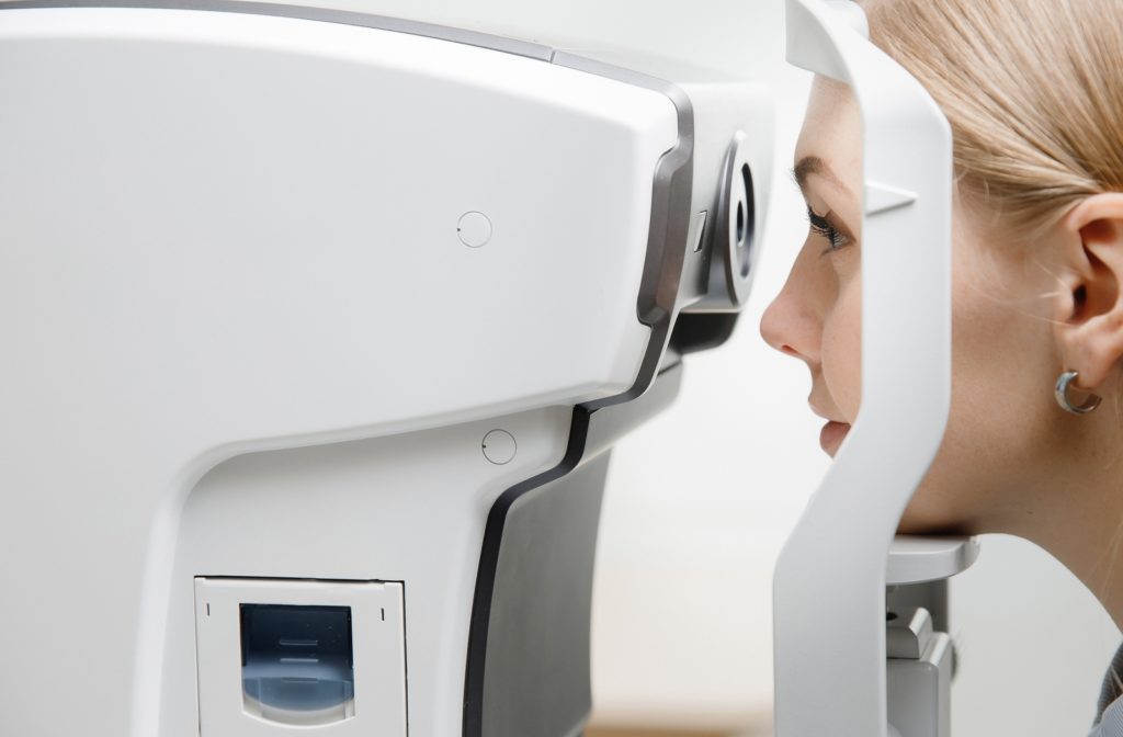
What Should You Expect During an OCT Test?
You may receive dilating eye drops before your exam begins. The OCT machine will scan your eyes for between 5 to 10 minutes while it creates your profile.
Your optometrist will look at these images when diagnosing any potential eye diseases. Afterward, they can recommend a customized treatment plan for your ocular needs.

What Diseases Can OCT Testing Help Diagnose?
OCT can help your optometrist diagnose several eye conditions, including:
- Age-related macular degeneration
- Glaucoma
- Diabetic retinopathy
- Macular hole
- Macular pucker
- Macular edema
- Central serous retinopathy
- Vitreous traction
OCT helps see changes in your optic nerve, helping to track disease progression.
Glaucoma
Glaucoma is one of the leading causes of blindness in adults over 60, caused by damage to your optic nerve. Most forms of this disease increase your intraocular pressure (IOP), leading to permanent vision loss. The common types of this disease include open-angle and angle-closure glaucoma.
While open-angle glaucoma can lead to blindness, it’s typically a gradually progressing condition. Angle-closure glaucoma can develop suddenly, leading to rapid vision loss. Visit your optometrist immediately if you experience:
- Severe headache
- Eye pain
- Nausea & vomiting
- Blurred vision
- Halos around lights
- Eye redness
Glaucoma doesn’t typically show symptoms until your vision is affected, but OCT testing can help identify damage to your optic nerve.
Age-Related Macular Degeneration
Age-related macular degeneration, known as AMD, is an eye disease affecting the central part of your retina. This area is called the macula, and it’s responsible for noticing fine details in your central vision.
Your macula can begin to wear down as you age, leading to vision loss. AMD comes in 2 forms, wet and dry.
Dry AMD occurs when small protein deposits called drusen develop. The light-sensitive cells in your macula begin to thin until they die. You may notice blind spots in your central vision over time.
Wet AMD is a rarer but more severe form of this disease. This condition causes new blood vessels to grow under the retina. These vessels can rupture, leaking blood and fluid into the eye.
Wet AMD is a medical emergency. You should see your eye doctor immediately if you experience changes in your central vision or lose the ability to see color and fine details.
Diabetic Retinopathy
Diabetic retinopathy is a form of diabetic eye disease where high blood sugar levels damage the blood vessels in your eyes. Damage causes the vessels to break and leak blood into your eye, causing the retinal tissue to swell and blur your vision. This is the early stage of this disease, known as nonproliferative diabetic retinopathy.
Abnormal blood vessels can begin to develop as diabetic retinopathy progresses. This stage of the disease is called proliferative diabetic retinopathy. It can lead to severe vision loss.
These are some of the common and significant eye diseases OCT can help diagnose. These conditions can lead to vision loss and affect your ocular health, but your optometrist can help identify signs of these conditions before they damage your eyes.
OCT Can Help Protect Your Vision
Optical coherence tomography testing can help diagnose potential eye diseases, but it’s only one tool in your optometrist’s arsenal. Your eye doctor can conduct several tests during an eye exam to help diagnose any potential problems. With regular eye exams, your optometrist can help protect your eyesight. Contact your optometrist if you need an eye exam or are experiencing any visual difficulties.